Why legacy pathology archives matter for rare-disease research
Why it matters
Rare-disease research needs larger, richer historical datasets
Digitized archives make decades of evidence searchable
Historical tissue can reveal patterns previously missed
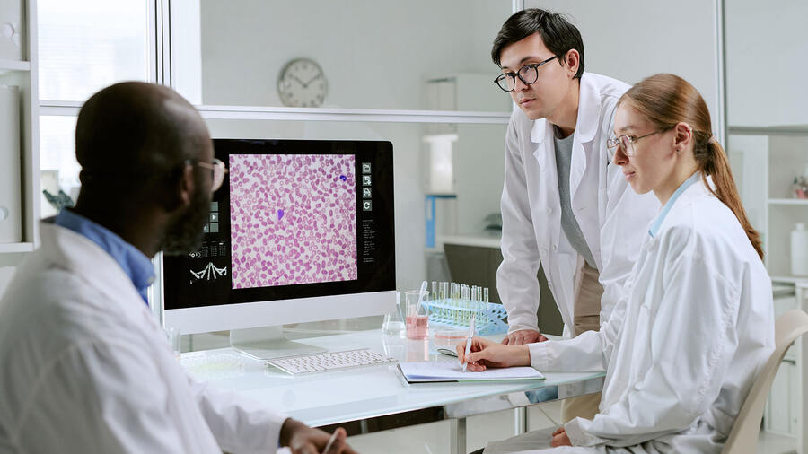
Pathology archives often contain decades of clinically valuable information that is difficult to access in its original format. Glass slides may reveal evidence of disease patterns and support diagnostic decisions and tissue characteristics that were essential at the time of creation, but as scientific research data they are difficult to search, share and study at scale.
As digital pathology matures, those archives are increasingly being viewed differently: not simply as storage, but as valuable research assets.
Archives hold more than history
This shift is significant because some of the most important questions in medicine cannot be answered with a small sample set. Rare diseases, unusual presentations and long-term retrospective analysis often require access to large collections of historical material. When slides are digitized, they become easier to review, compare, and organize for broader scientific use. Most importantly, not all slides need to be collocated for research as the data can be shared digitally. What was once a physical archive can begin to function as a structured digital dataset.
This is where ScanVan™ excels.
ScanVan is Leidos’ high-throughput whole-slide digitization platform, designed to bring scanning capability directly to the archive and help convert large volumes of glass slides into digital assets without requiring the collection to be moved off-site. In that sense, ScanVan is not the story itself; it is the operational example of how large-scale archive digitization can happen in practice.
The opportunity is impactful for hospital systems and pharmaceutical organizations that are interested in retrospective research, biomarker discovery and future AI-enabled analysis. Digital pathology makes it easier to examine archived tissue in a consistent format, identify patterns across cases and connect specimen-level information to the larger research environment. The larger and more organized the archive, the more useful it becomes for research teams trying to understand rare conditions, compare historical samples or generate hypotheses for future studies.
Unlocking value that’s already there
A platform like ScanVan supports high-volume digitization in a way that is practical for large, complex archives. The work still has to be done carefully—with strong attention to image quality, chain of custody and operational control—but once the archive is digitized, it becomes a resource that can be used in new ways.
In that sense, legacy pathology archives represent an untapped source of scientific value. They are already rich with information. Digitization simply makes that information easier to see, study and apply.
For health systems, the question is no longer whether the archive has value. The question is how to unlock it.
ScanVan by the numbers
Developed in collaboration with the University of Pittsburgh’s Computational Pathology and AI Center of Excellence, ScanVan is designed for collections ranging from thousands to millions of slides. Based on 12-hour scanning days, Monday through Friday, the platform can digitize:
- More than 16,000 slides per week
- More than 67,000 slides per month
- More than 800,000 slides per year
These figures demonstrate the operational scale required to turn a legacy archive into a usable research resource.
Three things to remember
- Digitized slides can be shared and studied without physically collocating pathology collections
- Archive digitization requires strong image quality, chain-of-custody and operational controls
ScanVan enables high-volume slide digitization while keeping pathology archives on-site